
Overview
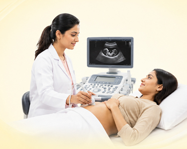
The Ultrasound Department at Lilavati Hospital, Gujarat, produces real-time images of internal structures using high-frequency sound waves. It doesn't use radiation as X-rays or CT scans do, so it's a safe choice for a lot of people, even pregnant women. A transducer is a small device that moves over the skin, sending sound waves into the body and picking up the echoes to make pictures. Ultrasound is especially good at imaging soft tissues, fluid-filled structures, and blood flow when used with Doppler technology. Most of the time, this procedure is done in an outpatient setting and doesn't take long.
The department of Ultrasound doesn't treat conditions, but it is very important for diagnosing them. It helps find cysts, tumors, gallstones, kidney stones, and fluid collections. In obstetrics, it is often used to monitor fetal development, and in gynecology, it is used to assess the health of reproductive organs. Doppler ultrasound assesses blood flow in vessels to detect blockages or circulation problems. It is also used to check for thyroid problems, soft-tissue lumps, and heart function (echocardiography).
Ultrasound is still one of the safest and easiest-to-use imaging tools in medicine. It can take pictures in real time, which makes it easy to make decisions and diagnose problems quickly. Ultrasound remains very important in both routine and specialized medical care, even as technology continues to improve, with higher resolution and portable devices.